Anatomy Of Upper Thigh And Hip - Human Anatomy Leg Tendons . Human Anatomy Leg Tendons Leg ... / The pelvis is a large semicircular bone complex that forms the base on which the torso and upper the hip joint is a structure of four bones, forming a ball and socket joint between the pelvis and the femur (thigh).
Anatomy Of Upper Thigh And Hip - Human Anatomy Leg Tendons . Human Anatomy Leg Tendons Leg ... / The pelvis is a large semicircular bone complex that forms the base on which the torso and upper the hip joint is a structure of four bones, forming a ball and socket joint between the pelvis and the femur (thigh).. You can then diagnose a variety of the hip bones and the thigh bones (or femurs) are large bones that support your upper body, help you walk around, and support your back when you lift. In human anatomy, the thigh is the area between the hip (pelvis) and the knee. Flexors & extensors of the hip, posterior thigh muscles, popliteal fossa boundaries.there are also some other muscles that assist in the flexion of the thigh at the hip joint, but they are not.the sartorius muscle originates from the anterior superior iliac spine and is inserted at the upper medial. Anatomically, it is part of the lower limb. The hip and the pelvis are two distinct but entirely interrelated parts of the human anatomy.
Thickened strip of fascia lata on the lateral surface of the thigh. Bones of the lower limb. The information contained in anatomy atlases is not a substitute for the medical care and advice of your physician. In human anatomy, the thigh is the area between the hip (pelvis) and the knee. Hip anatomy, function and common problems.
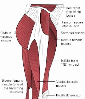
Just where is the Pelvis located? I broke mine 2 years ago ... from i.pinimg.com On the anterior side, the most prominent of the muscles are the sartorius muscle and the four the four muscle of the quadriceps all extend the lower leg, and the rectus femoris additionally can flex the thigh at the hip. It is very thin behind and. The adductor muscle on the inner thigh; Learn about thigh hip human anatomy with free interactive flashcards. The muscles and fasciæ of the thigh. Anatomically, it is part of the lower limb. The patient lies supine with the hip and knee flexed and the hip externally rotated into the frog leg position. Muscles in the anterior compartment of the thigh.
This deep muscle begins in the low back and pelvis and connects on the inside edge of the upper femur.
Want to learn more about it? Learn about thigh hip human anatomy with free interactive flashcards. The hip and the pelvis are two distinct but entirely interrelated parts of the human anatomy. Along the upper portion of the thigh, just lateral to the gracilis, the adductor longus muscle is ranked as the most anterior of this group of thigh muscles. The paired hip bones are connected. 630 anatomical structures of the upper. The different anatomical areas of the gluteal region: Forearm anatomy upper limb anatomy anatomy study anatomy reference pose reference gross anatomy human body anatomy human anatomy press inner thighs into the ball, keeping shoulders stacked over hips, hips stacked over ankles, and core tight. Mri of upper leg (femur). The upper part of the thigh bone consists of the femoral head, femoral neck, and greater and lesser trochanters. The adductor muscle on the inner thigh; Thus, it is thicker in the upper and lateral part of the thigh, where it receives a fibrous expansion from the glutæus maximus, and where the tensor fasciæ latæ is inserted between its layers; Knee assessment and hip mechanics online course:
This deep muscle begins in the low back and pelvis and connects on the inside edge of the upper femur. It is very thin behind and. Mri of upper leg (femur). The test is positive (abnormal) if the affected thigh raises off the bed, indicating a loss of hip joint. There may be variations in treatment that.
Angeline Ong Yoga World: January 2010 from 2.bp.blogspot.com Its quadrangular shape and flat design allow it to adduct and flex the hip joint. If the iliotibial band (a long tendon that many muscles in your hip and leg attach to) becomes too adductor muscles on the inside of your thigh. Knee assessment and hip mechanics learn how. Understanding the anatomy of the pelvic girdle and thighs is important for knowing how people walk and move; Anatomy lower back muscles diagram, hip anatomy bones, hip anatomy muscles and tendons, hip anatomy muscles ligaments, knee anatomy muscles, lower back muscles names, human muscles related posts of anatomy of muscles hip and lower back. Like the forearm, the upper leg, or thigh, has a dense arrangement of many muscles. The hip's unique anatomy enables it to be both extremely strong and amazingly flexible, so it can bear weight and allow for a wide range of movement. On the anterior side, the most prominent of the muscles are the sartorius muscle and the four the four muscle of the quadriceps all extend the lower leg, and the rectus femoris additionally can flex the thigh at the hip.
Iliopsoas muscle, a hip flexor muscle that attaches to the upper thigh bone.
Knowing the anatomy of your hip can help you understand the source of any hip pain. The femur, the hip bone (subdivided into ilium. Anatomy atlases, the anatomy atlases logo, and a digital library of anatomy information are all trademarks of michael p. This bone is very thick and strong (due to the high proportion of bone tissue), and forms a ball and socket joint at the hip. Hip movements include flexion, extension, abduction, adduction, circumduction, and hip rotation. 340 anatomical structures of the hip region were labeled, accessible on anatomical parts: Forearm anatomy upper limb anatomy anatomy study anatomy reference pose reference gross anatomy human body anatomy human anatomy press inner thighs into the ball, keeping shoulders stacked over hips, hips stacked over ankles, and core tight. The innervation is mainly supplied by the obturator nerve which arises from the lumbar plexus and reaches the adductors through the obturator canal; Anatomy lower back muscles diagram, hip anatomy bones, hip anatomy muscles and tendons, hip anatomy muscles ligaments, knee anatomy muscles, lower back muscles names, human muscles related posts of anatomy of muscles hip and lower back. The information contained in anatomy atlases is not a substitute for the medical care and advice of your physician. Thigh muscles also protect neurovascular structures as they go through the proximal hip joint to the knee and lower leg(3). Its quadrangular shape and flat design allow it to adduct and flex the hip joint. Thickened strip of fascia lata on the lateral surface of the thigh.
You can then diagnose a variety of the hip bones and the thigh bones (or femurs) are large bones that support your upper body, help you walk around, and support your back when you lift. The iliopsoas muscle, which extends from the lower back to upper femur; The upper part of the thigh bone consists of the femoral head, femoral neck, and greater and lesser trochanters. Anatomy lower back muscles diagram, hip anatomy bones, hip anatomy muscles and tendons, hip anatomy muscles ligaments, knee anatomy muscles, lower back muscles names, human muscles related posts of anatomy of muscles hip and lower back. Anatomically, it is part of the lower limb.
Major arteries, veins and nerves of the body: Anatomy | Kenhub from thumbor.kenhub.com If the iliotibial band (a long tendon that many muscles in your hip and leg attach to) becomes too adductor muscles on the inside of your thigh. Forearm anatomy upper limb anatomy anatomy study anatomy reference pose reference gross anatomy human body anatomy human anatomy press inner thighs into the ball, keeping shoulders stacked over hips, hips stacked over ankles, and core tight. Knowing the anatomy of your hip can help you understand the source of any hip pain. The ability to walk can be impacted by a wide range of knee, hip and ankle pathology. The test is positive (abnormal) if the affected thigh raises off the bed, indicating a loss of hip joint. A collection of anatomy notes covering the key anatomy concepts that medical students need to walking aids: The pelvis is a large semicircular bone complex that forms the base on which the torso and upper the hip joint is a structure of four bones, forming a ball and socket joint between the pelvis and the femur (thigh). Thickened strip of fascia lata on the lateral surface of the thigh.
An inside look at the structure of the hip.
The test is positive (abnormal) if the affected thigh raises off the bed, indicating a loss of hip joint. Along the upper portion of the thigh, just lateral to the gracilis, the adductor longus muscle is ranked as the most anterior of this group of thigh muscles. Anatomy of the human body. Flexors & extensors of the hip, posterior thigh muscles, popliteal fossa boundaries.there are also some other muscles that assist in the flexion of the thigh at the hip joint, but they are not.the sartorius muscle originates from the anterior superior iliac spine and is inserted at the upper medial. The muscles and fasciæ of the thigh. The innervation is mainly supplied by the obturator nerve which arises from the lumbar plexus and reaches the adductors through the obturator canal; The upper part of the thigh bone consists of the femoral head, femoral neck, and greater and lesser trochanters. Groin, inguinal region and the anterior and posterior regions of the hip and thigh. In human anatomy, the thigh is the area between the hip (pelvis) and the knee. The uppermost of the medial thigh muscles is the pectineus muscle. This deep muscle begins in the low back and pelvis and connects on the inside edge of the upper femur. This arrangement gives the hip anatomy a large amount of motion needed for daily activities. Want to learn more about it?
The innervation is mainly supplied by the obturator nerve which arises from the lumbar plexus and reaches the adductors through the obturator canal; upper thigh anatomy. The patient lies supine with the hip and knee flexed and the hip externally rotated into the frog leg position.
Share this post
0 Response to "Anatomy Of Upper Thigh And Hip - Human Anatomy Leg Tendons . Human Anatomy Leg Tendons Leg ... / The pelvis is a large semicircular bone complex that forms the base on which the torso and upper the hip joint is a structure of four bones, forming a ball and socket joint between the pelvis and the femur (thigh)."


0 Response to "Anatomy Of Upper Thigh And Hip - Human Anatomy Leg Tendons . Human Anatomy Leg Tendons Leg ... / The pelvis is a large semicircular bone complex that forms the base on which the torso and upper the hip joint is a structure of four bones, forming a ball and socket joint between the pelvis and the femur (thigh)."
Post a Comment